What are the dark moles on my dog s belly

Hyperpigmentation (Acanthosis Nigricans) in Dogs
Hyperpigmentation is a darkening and thickening of the skin seen in dogs. It is not a specific disease but a reaction of a dogs body to certain conditions.
Hyperpigmentation appears as light-brown-to-black, velvety, rough areas of thickened, often hairless skin. The usual sites are in the legs and groin area. It can be primary or secondary. Primary diseases that cause hyperpigmentation can occur in any breed but especially Dachshunds. Signs are usually evident by 1 year of age. Secondary hyperpigmentation is relatively common and can occur in any breed of dog, most commonly those breeds prone to obesity, hormonal abnormalities, allergies, contact dermatitis, and skin infections. Secondary hyperpigmentation is triggered by inflammation and/or friction. Inflammation leads to additional skin changes, such as thickened skin, hair loss, odor, and pain.
The edges of inflamed areas are often red, a sign of secondary bacterial or yeast infection. With time, it may spread to the lower neck, groin, abdomen, hocks, eyes, ears, and the area between the anus and the external genital organs. Itching is variable. When it occurs, it may be caused by the underlying disease or by a secondary infection. As the condition progresses, secondary hair loss, fluid discharge, and infections develop.
Diagnosis is by appearance of signs on the animal. In a young Dachshund, your veterinarian will want to eliminate other causes of the signs. A careful history and physical examination will be performed to identify an underlying cause. The presence of secondary hyperpigmentation always suggests an underlying disease. Skin scrapings are taken to exclude other causes (parasites, for example), especially in young dogs. Impression smears are used to identify bacterial infections. Depending on other signs, endocrine function tests for thyroid and adrenal disease may be used to check for underlying hormonal abnormalities. Skin testing, a food trial, or both may be necessary to test for allergies. Skin biopsies may be made to check for a condition called seborrhea. In most cases, your veterinarian will want to treat any secondary bacterial infections before proceeding with other diagnostic tests.
Primary hyperpigmentation in Dachshunds is not curable. In some dogs, the condition is only cosmetic and does not require treatment. If inflammation is present, early cases may respond to shampoo treatment and steroid ointments. As signs progress, other treatment, such as medication given by mouth or injection, may be useful. The concurrent treatment of secondary infections is helpful and is required before steroids are administered. Medicated shampoos are often beneficial for removing excess oil and odor but must be used regularly.
In secondary hyperpigmentation, the affected areas will go away on their own after identification and treatment of the underlying cause. However, this will not occur if secondary bacterial and yeast infections are not treated and controlled. Many affected dogs benefit greatly from appropriate antibiotics and medicated shampoos (2 to 3 times per week). Thus, many veterinarians will prescribe such treatments. Owners need to be patient with these treatment programs. The signs of hyperpigmentation resolve slowly; it may take months for the dogs skin to return to normal.
Also see professional content regarding acanthosis nigricans.
6 Types of Moles on Dogs [With Pics] and What to Do
This article was updated on October 16th, 2023
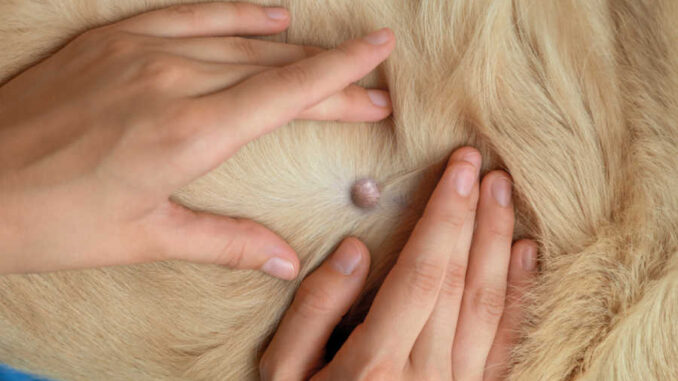
When an owner comes to my clinic because they are worried about a new growth or mole on their dog, the question on everyones lips is, What is it?.
When a new skin lesion resembles a mole, owners will inevitably wonder: do dogs have moles? The answer is yes. However, we refer to them as nevus (singular) or nevi (plural). Some owners and vets, however, will use the word mole interchangeably with skin tag or wart.
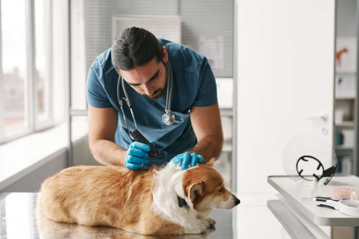
Types of moles (with pictures)
Technically, a mole is a nevus. This is a benign skin growth that is usually small and quite symmetrical. Uncommonly, a nevus can be cancerous or can transform into a cancer. Cancerous moles on dogs are quite rare, thankfully.
As mentioned, other types of lesions such as a cyst, skin tag or wart may also be referred to as moles by some people. Lets take a look at some of the more common types of moles or bumps in dogs, with pictures:
Skin Tags
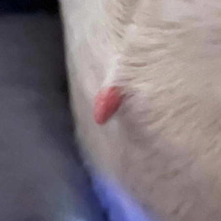
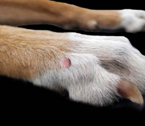
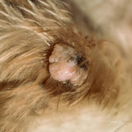
In older dogs especially, skin tags start to appear. We often see them on the face, elbows, armpits, and ankles. They can dangle from the skin and may be dark or a fleshy pink color. They are slow-growing and should not bother the dog.
Generally, we would monitor skin tags but would not remove them unless they were becoming a nuisance. Learn more about Skin Tags.
Cysts
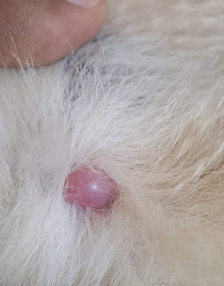
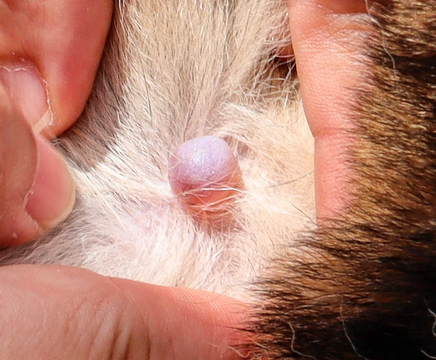
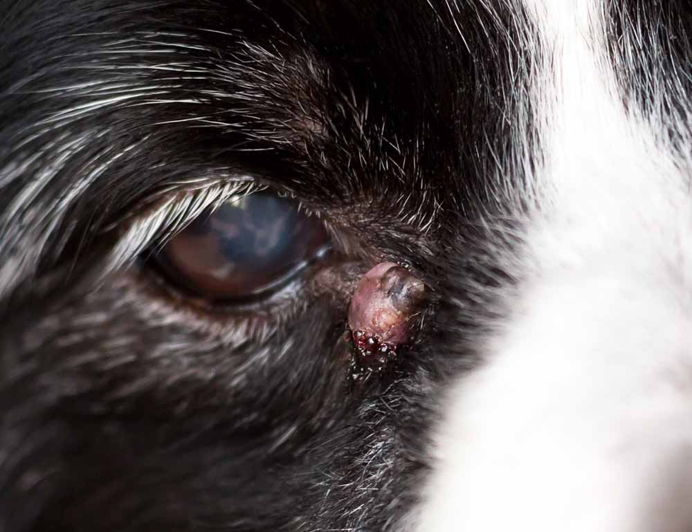
Dogs develop all kinds of cysts, some of which are fluid-filled and others that contain a thicker substance that resembles cottage cheese.
Canine cysts can appear anywhere on the body and may grow to a substantial size. However, most cysts remain quite small. If a cyst is drained or ruptures, it will refill quickly. While cysts usually do not need to be removed, removing one involves a surgical procedure to extract the cyst wall. Learn more about Cysts in Dogs.
Warts or sebaceous adenomas
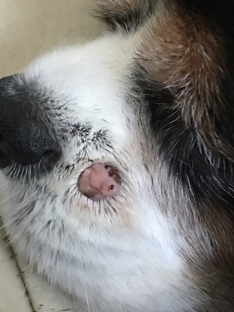
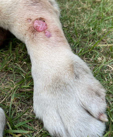
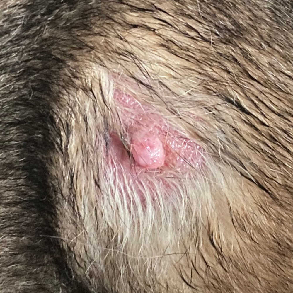
Warts tend to be light pink and can resemble small cauliflowers or brains. We tend to see warts in two dog populations: puppies and seniors. This is because both of these age groups struggle to fight off infections and warts are spread by a virus. For younger dogs, warts usually resolve within a few months. For older dogs, they may persist and grow slowly over time. Learn more about Warts in Dogs with pictures and veterinarian info.
Cancer
Cancerous lumps come in all shapes and sizes, as shown in the pictures below.
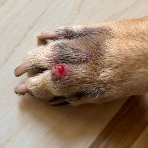
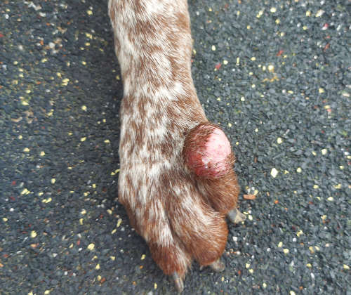
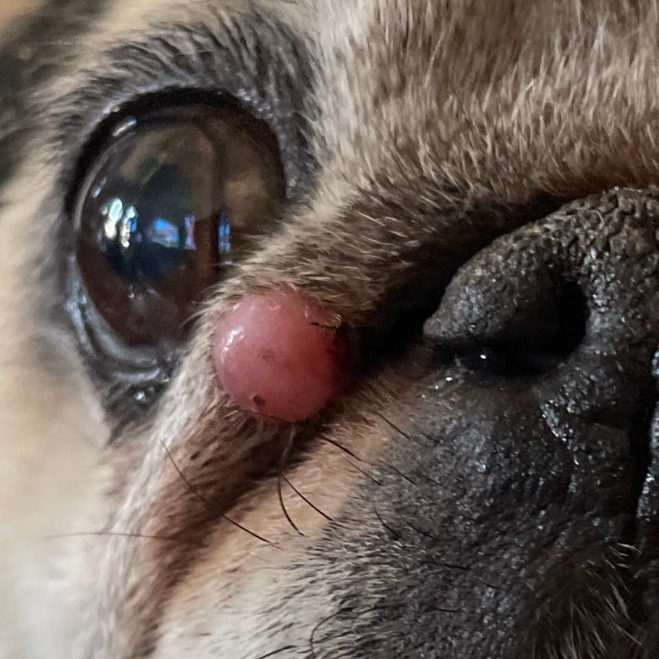
A black mole on a dog (rather than white, pink or red) is more likely to be a cancer called melanoma. Some of the more common places where malignant melanoma occurs include the mouth, near the claws, and sometimes within the eye. Learn more about Cancerous Lumps.
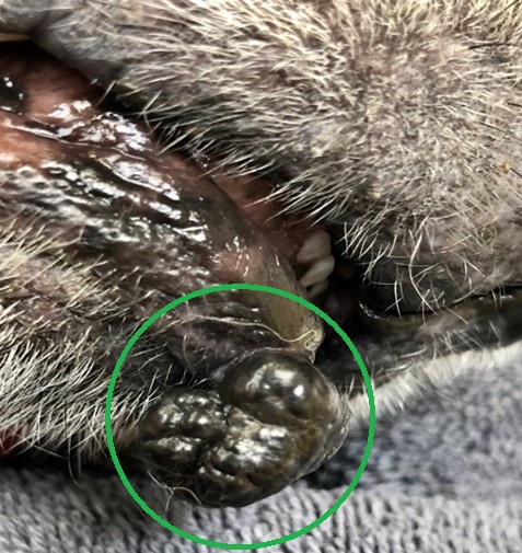
True nevi
A small black or brown mole on a dog may be a true nevus. These moles can appear on the face, flank, and paws, as well as other locations. As these lesions are benign, there is generally no need to intervene or provide treatment. However, a vet may discuss sampling the lesion to rule out anything more sinister, such as a melanoma.
Related post: Pictures of Common Lumps & Bumps on Dogs.
What to do when you find a mole or bump on your dog
1. Take pictures: When a new bump is noticed, it is a good idea to snap a photo, so you can assess if it is growing or changing over the coming days and weeks.
2. Consider a buster collar: If your dog has been licking or chewing at it, it is wise to use a buster collar to prevent this. This helps ensure the lesion does not become infected.
3. Reach out to your vet: A lesion that is not going away after a few days should be examined by a vet. Most of the time, new moles or warts will be something we monitor. Less often, your vet may discuss sampling or removal of the lump, particularly if they are concerned about cancerous tumors such as melanomas or mast cell tumors.
When is it ok to wait-&-see?
If youve only just noticed a small lesion and it is not bothering your dog, you may monitor it over a few days. It could be a small scab or insect sting, which will resolve quickly.
It is sensible to consult your veterinarian if the new lesion is persisting for over a week or if it seems to be quickly growing and changing. A consultation should also be booked if a lesion suddenly gets bigger or becomes infected.
Veterinary treatment, costs, and recovery
For many new lumps and bumps, the cost involved may be limited to the consult (about $60-100), as the vet may feel that the lesion is likely benign and needs no intervention. However, it is important to remember that the only way to know this is to sample and test the lesion.
If the mole needs to be investigated, this will be more costly. This can involve a Fine Needle Aspirate ($150-250) or biopsy ($200-400).
Removing a mole may be more costly, as it usually means the pet needs to be under an anesthetic. This can mean a bill of $400-900. This price should include the lab fees for analyzing the growth.
Frequently Asked Questions
What causes the growth of moles or bumps in dogs?
There is likely a large genetic component when it comes to the development of these lesions, but other factors can play a role, too.
- Exposure to sun. Dogs who spend a lot of time in the sun, particularly those who are short-furred, seem more prone to moles and skin tags.
- Weight gain. Dogs who carry more fat than they should can be more prone to developing certain lesions.
- Friction. When skin rubs against itself, especially in places like the armpits and groin, this can lead to skin tags developing.
Can moles or bumps on dogs be cancerous?
Yes, it is possible for these skin lesions to be malignant, which is why a vet visit should be booked for any lesion that is not going away quickly. Read our article about cancerous lesions and bumps in dogs.
How can I differentiate between a benign and malignant mole or bump?
There is no definitive way to do this without sampling the lesion and looking at it under the microscope. However, malignant lesions do share certain characteristics, such as being quick to grow, ulcerating, and becoming infected. Learn more: cancerous lesions and bumps.
Can moles or bumps on one dog spread to other dogs or humans?
No, moles cannot spread and are not contagious. Warts (papillomas), on the other hand, can spread from dog to dog.
Are certain dog breeds more prone to developing bumps?
Yes, there is a genetic component, so we will see certain lumps more often in certain breeds. Poodles, Schnauzers, and Golden Retrievers may be over-represented.
Dr Linda Simon (MVB MRCVS) has 10 years of experience as a veterinarian. She is a veterinary surgeon with a special interest in geriatric patient care, dermatology and endocrinology. She is a member of the British Royal College of Veterinary Surgeons. She graduated top of her class from UCD School of Veterinary Medicine in Dublin in 2013. Linda has also worked as a locum vet in a range of clinics, including 24 hour emergency clinics and busy charity clinics.
View all posts
Disclaimer: This website's content is not a substitute for veterinary care. Always consult with your veterinarian for healthcare decisions. Read More.
Cancerous Moles on Dogs
Cancerous moles may develop on the surface of the skin. Skin cancer is more common in senior dogs and dogs that have light colored coats. The moles may occur due to an uncontrollable development of cells. Not all moles are cancerous, but your dog should be checked by a specialist if he has any moles or abnormal skin growths.
Moles on Dogs
Moles on dogs may be benign or malignant and develop due to an abnormal division and multiplication of the cells. The division and multiplication of cells is controlled by the genes in the nucleus of cells. However, there may be some external factors that could influence the abnormal growth of skin cells.
Appearance of Canine Cancerous Moles
Moles are hard lumps and in dogs, they are typically dark in color. The moles may be present on any area of the dogs skin. Many dogs have moles, but not all of these are cancerous. Typically, moles that grow at a fast rate are cancerous. There are a few means that you could suspect the moles on your pet are cancerous:
- Cancerous moles are dark and grow on skin areas with hair (the color of the moles may change)
- The moles may have various sizes, but the edges are irregular
- Are raised and may bleed occasionally, as there are many blood vessels in the area
Benign moles may sometimes look like cancerous ones, so its always a good idea to have a veterinary checkup and have a clear diagnosis.
Types of Cancerous Moles on Dogs
There are several types of cancerous moles including:
- Melanoma, which is made up of malignant cells that typically develop in an older mole and affects certain breeds more often: Cocker Spaniels, Scottish Terrier and Boston Terriers.
- Sebaceous adenomas, which are lumps affecting the sebaceous glands. These moles are light in color and are smaller. Cocker Spaniels seem to be more often affected by this type of skin cancer
- Skin carcinomas have the appearance of cauliflower and are most frequently located on feet and legs
- Mast cell tumors, which often affect the legs or the abdominal area. This type of cancer is more common in Boxers
Diagnosing Cancerous Moles
A sample of cells is required to be biopsied to see if the mole is benign or malignant. The pathologist can establish the type of cancer and the stage of the cancer.
Other tests are needed if the cancer is suspected to be in a more advanced stage and it is present in neighboring lymph nodes or other organs.
Treatment Options for Cancerous Moles
Cancerous moles on dogs may be fully treated, but a timely detection is essential. Surgical excision is possible in the early stages of the disease and in some dogs, the moles will not be recurrent. If the cancer affects other organs, surgery is not an effective course of treatment and chemotherapy will be recommended.


