What are the little black growths on my dogs skin

6 Types of Black Lumps on Dogs [With Pictures]
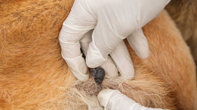
This article was updated on October 18th, 2023

It can be concerning to see a new black lump on your dog, particularly because black growths may mean cancer in human medicine. When I worked as a veterinarian, I encountered various causes for black bumps on dogs: some were cancerous, but others were benign and nothing to worry about. Lets look at the most common types with pictures selected by our veterinarians.
Black lumps, bumps, or growths on dogs
1. Warts or adenomas
Very common, mostly in senior dogsWarts, caused by canine papillomavirus, and adenomas are some of the most common types of small lumps on dogs. These masses are benign, irregularly shaped like cauliflower, and usually pale in appearance. Below are two examples of black warts. Notice the irregular, raised shapes:
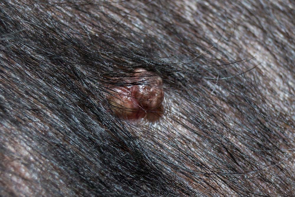
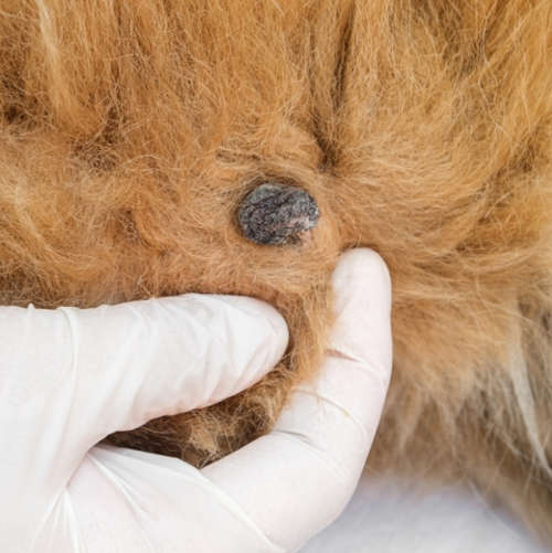
Although warts and adenomas are not a major concern, they can become infected or ulcerated. When warts become inflamed or infected, blood or pigment can turn the bumps dark. When warts become problematic, the treatment of choice is surgical removal. Learn more about Warts (Pictures & Treatments).
2. Skin tags
Very commonSkin tags are benign fibrous growths that usually occur in high-friction areas on your dogs skin. With a variable appearance, tags may look like raised bumps or attach to the body on a narrow stalk. They appear with or without hair. Skin tags are very common in dogs and can sometimes be black.
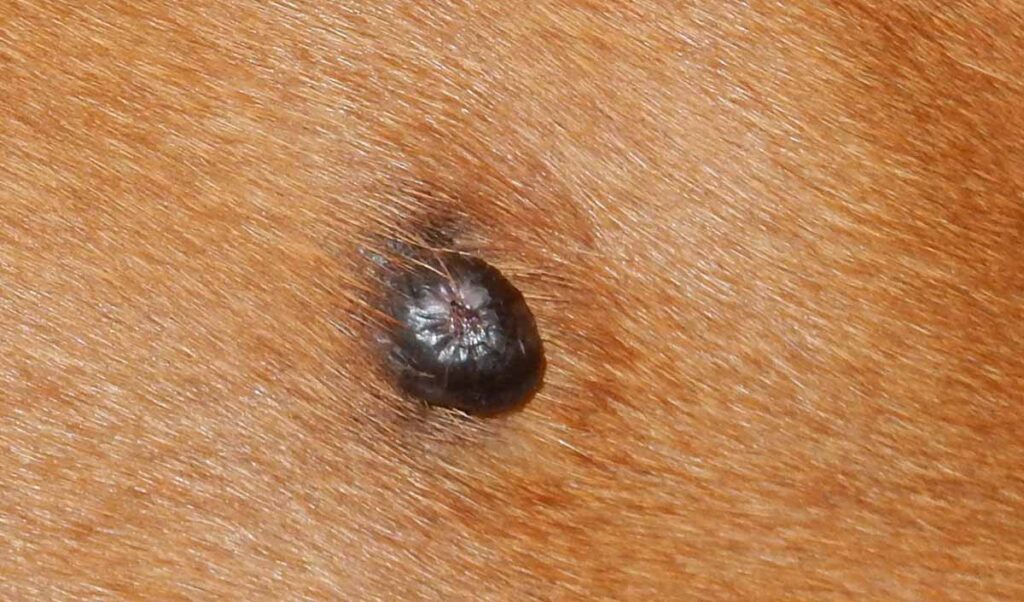
You may suddenly see a new skin tag on your dog, but they usually take time to develop. They are generally harmless but can become irritated and painful from constant rubbing. As long as your dog isnt bothered, skin tags can be left alone. You should also contact your veterinarian if you notice any changes in color, shape, or size. Learn more about Skin Tags: Pictures, Diagnosis, Treatment.
3. Histiocytomas turning black over time
Very common, mostly in younger dogs (up to 3 years old)Histiocytomas are benign, dome-shaped lumps that arise when immune cells (histiocytes) overgrow. They suddenly appear on the face, ear flaps, or legs, can grow rapidly, and may ulcerate. Although histiocytomas are normally red/pink in color, they can turn black over time, as shown on the picture below:
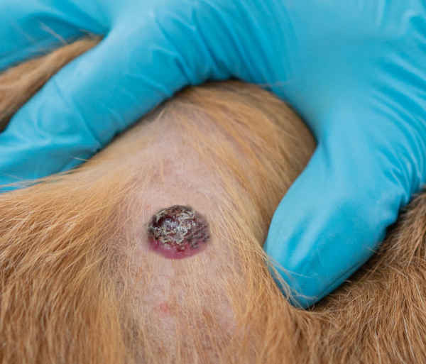
If you have a young dog that develops a histiocytoma, discourage him from licking, chewing, or scratching the bump. This will help to prevent inflammation, infection, and ulceration.
Most histiocytomas will resolve spontaneously in 1-3 months without treatment. Learn more about Histiocytomas.
4. Cancerous tumors: melanomas
Melanomas are malignant growths that appear as black or dark lumps, often found in older dogs on their lips or mouth (as shown on the images below). When pigment-producing cells known as melanocytes grow proliferatively, they usually create brown or black lumps because the cells contain melanin granules.
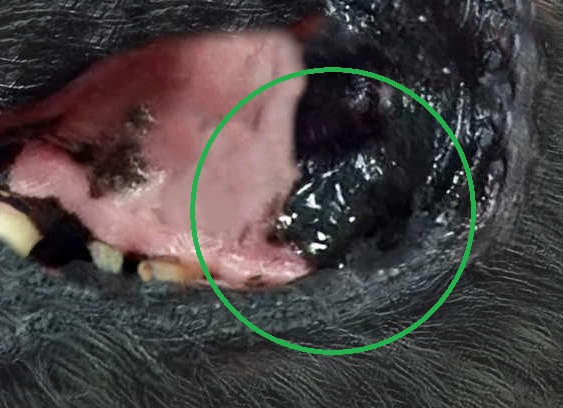
These tumors often grow rapidly and spread quickly to other parts of the body.
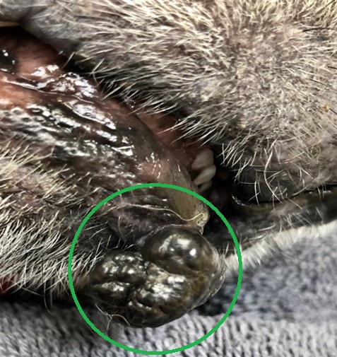
Black or dark cancerous lumps:
Cancerous lumps tend to grow quickly and in an unpredictable manner, which often results in an irregular shape. This is illustrated by the black lumps in the two pictures below:
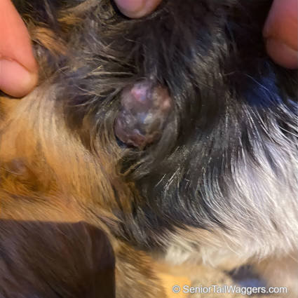
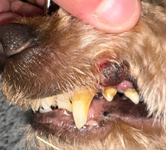
Early diagnosis and treatment are important because they significantly impact your dogs diagnosis. The primary method to treat melanomas is the surgical removal of the tumor along with surrounding tissues that may be affected. Learn more about Melanomas (Pictures & Treatments).
If a lump or lesion appears red, black, or generally unhealthy in appearance, it may be more likely to be malignant. Cancerous lesions and lumps are also often harder and firmer to the touch.However, keep in mind that it is generally not possible to tell if a lump is cancerous or not just by looking at it: a biopsy is generally needed (more information regarding this is provided later in this article).
5. Darker ticks
Some embedded ticks can mimic black bumps when you see them on your dogs skin. Depending on where you live, some common ticks on dogs include deer ticks, American dog ticks, and Lone Star ticks. If you find an embedded tick on your dog, you may remove it at home or have your veterinarian extract the parasite.
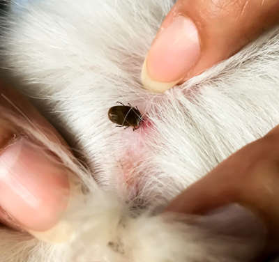
To remove a tick, use fine-tipped tweezers and grasp the pest as close to your dogs skin as possible. Pull the tick straight out with gentle, steady pressure. Avoid twisting, as you may break off the head, which can cause infection. Once the tick is out, examine it to make sure you have the entire body, then disinfect/wash the bite area.
6. Hygromas
Hygromas typically form on the bony prominences of large dogs who lie on hard surfaces like pavement or tiles. They form in an effort to protect the dogs skeleton and to minimize friction. In the photo below, the dog has a hygroma on the elbow and the fur above it is missing; this would be quite typical:
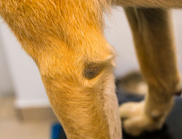
Hygromas
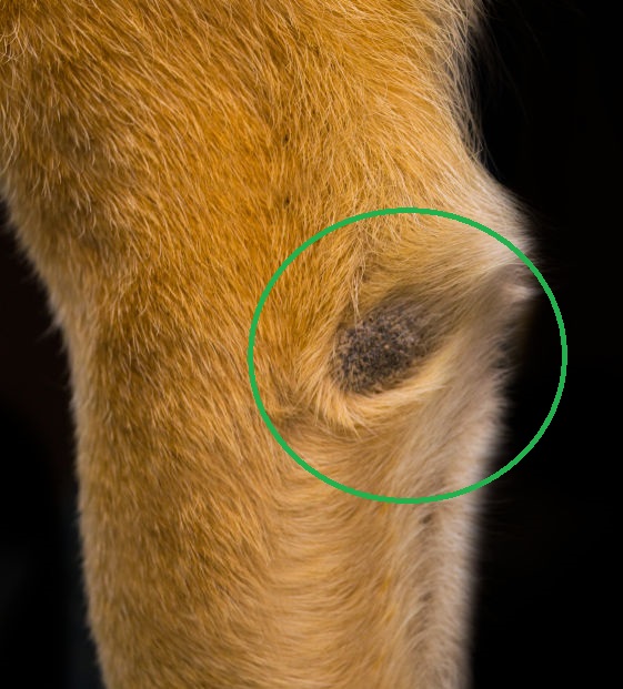
A biopsy is generally needed to confirm the nature of a lump
When owners bring their dogs to our clinic because of a black lump, we typically perform a physical examination and run tests that may include:
- evaluating the characteristics of the mass including size, shape, depth, and more
- taking a fine needle aspirate or biopsy and submitting it to the lab for analysis
- running bloodwork to check for endocrine disorders or signs of infection
- skin scrapings or hair samples if we suspect issues such as mites or skin infections
- impression smears to help diagnose the cause of scabs
Estimated cost of diagnosis
Depending on your dogs particular situation, your vet may want to charge the following:
- office visit and initial exam approximately $50-100
- bloodwork $150-300 in most cases
- aspirate, skin scrapings, or impression smears with cytology $25-250
- biopsy with pathology $300-600
- bacterial or fungal cultures $300-400
Why are some lumps black?
Bumps or growths on dogs can be black mostly because they are one of the following:
- blood-filled lumps: when a dogs skin experiences trauma, blood may collect at the point of injury. Over time, the blood dries and turns reddish-brown or black, like a scab.
- tumors: whether the growth is benign or malignant, it may collect pigment granules known as melanin that turn the bump black. They often appear in response to skin inflammation, but the cells that produce melanin sometimes turn cancerous, causing malignant black tumors (known as melanomas).
Top causes of black spots or scabs on the skin
Below, well look at various reasons that black spots, scabs, or raised, itchy patches of dark skin may suddenly appear on your dog.
1. Hyperpigmentation
When your dogs skin is traumatized, melanocytes produce extra pigmentation to protect the damaged areas resulting in black spots on your dogs skin. Common causes of hyperpigmentation include injuries, allergies, parasites, aging changes, cancer, or endocrine disorders like Cushings disease or hypothyroidism.
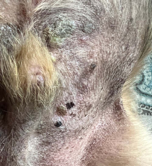
If your senior dog develops black patches or spots on his skin but there arent any other changes, its probably age spots. However, you should contact your veterinarian if there are other changes to the skin like:
- scaliness
- thickening
- roughening
- itchiness
- crustiness
- redness around the margins
These symptoms often indicate an underlying condition that requires treatment.
2. Injury
Injuries such as scrapes or cuts are the most common cause of black scabs in dogs. When the skin surface is traumatized, it seeps blood and platelets. A clot forms and dries to form a protective covering over the skin. Over time, the scab turns dark brown to black.
As long as the scab doesnt turn yellow or become infected, and your dog leaves it alone, you can let the skin heal naturally. If your furbaby continually licks and abrades the area, you may need to use an E-collar to protect the wound from further irritation.
3. Allergies
When your dog reacts to a food-based or environmental allergen, he can develop an allergy skin rash or hives. At first, the skin will be red and irritated, but if the inflammation is chronic, the area may become thickened, darkly pigmented, and form black or dark scabs from incessant itching.
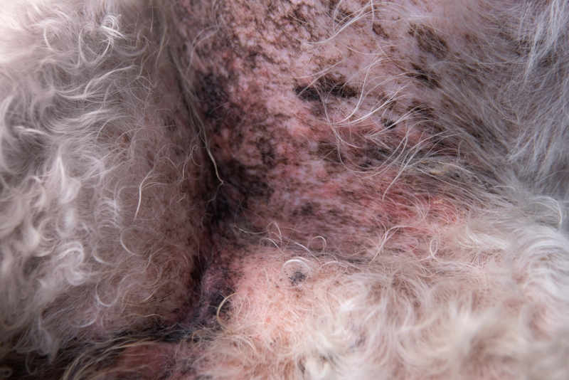
If your dog has chronic allergies, consult with your veterinarian. He may benefit from steroid treatment or a hypoallergenic diet.
4. Parasites
External parasites like fleas and mites can cause black scabs or spots on your dog. Fleas usually jump on your dog to feed and often leave behind flea dire (feces) that resemble tiny black scabs. Additionally, their bites can trigger an allergic reaction known as flea bite dermatitis that can leave black scabs.
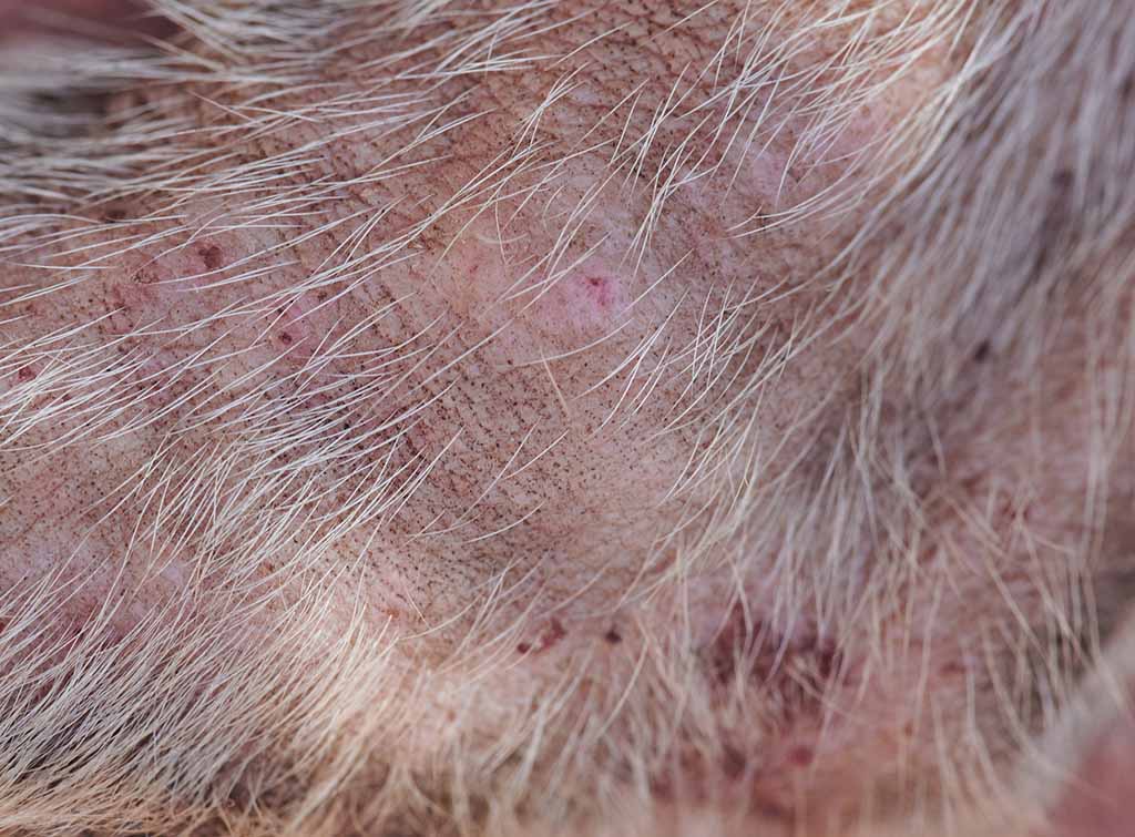
On the other hand, mites tend to burrow under your dogs skin, causing inflammation and irritation. As a result, your pup will scratch or chew the area continuously and cause skin trauma and scabbing.
To prevent skin issues from external parasites, keep your dog up-to-date on preventative treatments for parasites. If you suspect that your dog has mites or fleas, contact your veterinarian.
5. Skin infections
Skin infections in dogs are usually caused by Staphylococcus spp. bacteria or fungal infections(most commonly ringworm or yeast). These conditions irritate the skin and cause inflammation that can lead to scabbing or hyperpigmentation.
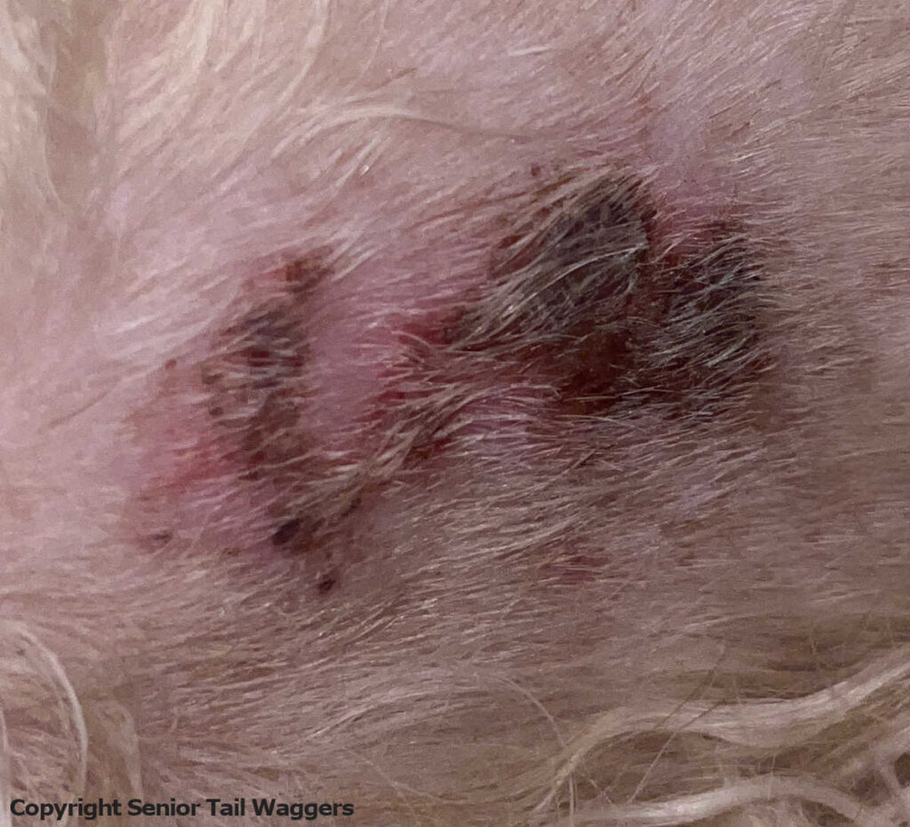
Skin infections should be examined by your veterinarian to properly diagnose the underlying cause and administer the appropriate treatments. Depending on the type and severity of the infection, your veterinarian may prescribe topical treatments, antifungal medication, or antibiotics.
Learn more about black spots on dog skin or black scabs on dogs (with more pictures and advice from our veterinarian team).
Learn more about lumps and bumps on dogs:
Dr. Liz (Elizabeth) Guise graduated from the University of Minnesota with a doctorate in Veterinary Medicine (DVM). She worked as a veterinarian for two years before working for the US Department of Agriculture for 13 years.
View all posts
Disclaimer: This website's content is not a substitute for veterinary care. Always consult with your veterinarian for healthcare decisions. Read More.
Lumps, Bumps, and Cysts on Dogs
Types of Lumps and Bumps on Dogs
Here are several common skin growths found in dogs, along with info on what they look like and what to watch for:
Benign Tumors
Tumors that are benign are not invasive or likely to spread to other body areas.
Histiocytoma
A histiocytoma is a benign skin growth that usually occurs in dogs less than 2 years of age. They are found on the front half of a dogs body, usually on the head or legs. Rarely, they can be seen in older dogs or on other areas of the body.
Histiocytomas are pink and fleshy but may get bigger and seem more irritated before improving. These tumors usually regress spontaneously over time without treatment and arise from the skins immune cells. They can be diagnosed through microscopic examination of a sample of cells from the growth.
Lipoma
A lipoma may show up anywhere on a dogs body but is common on the trunk and legs. Lipomas come from fat cells under the skin or are found in muscle tissue. They usually develop in older, overweight dogs. They may become quite large or appear in multiple locations.
A vet can diagnose a lipoma by taking a small sample of cells from the growth to look for fat droplets. No treatment is needed, but these should be monitored for rapid changes. They will gradually enlarge with time, and may bother your dog if theyre located in an area that interferes with motion. When lipomas start to bother your pet, you can consider surgical removal.
Papilloma
A papilloma in young dogs is a contagious, wart-like growth that usually occurs in and around the mouth. In older dogs, they might be seen around the eyes or on other areas of the body. Papillomas are caused by a virus that can be spread through direct contact with an infected dog or contaminated items, such as toys or feeding bowls.
They appear small, fleshy, and round with a cauliflower-like texture to the surface. Many will dry up and fall off within a few months as the dogs immune system matures. Severe cases may make eating or swallowing difficult and require treatment by surgical removal. Medications and other treatment methods are also available, including crushing of the warts to stimulate the immune system.
Another type of papilloma is a skin wart that is more common in older dogs. These are usually solitary and not caused by a virus. These bumps may have a hardened surface that looks like a cauliflower. An inverted papilloma may also be seen in young adult dogs, especially on the lower abdomen. If the growth bothers the pet, surgical removal is an option.
Skin Tag
A skin tag grows in places where a dogs skin rubs together. They are overgrowths of the connective tissue in the skin. They are the same color as the skin but extend out from the surface on thin stalks.
Skin tags are common in older dogs and certain breeds. No treatment is needed, but these can be surgically removed if they are bothersome.
Sebaceous Gland Tumor
A sebaceous gland tumor is commonly found in older dogs. They are typically smaller than a pea and may develop in any location. Some will bleed or secrete a material that forms a crust. Large breeds often form these on their head, specifically their eyelids, and they may be black in color. Treatment is not necessary, but surgical removal may be considered when the growth is bothersome.
Meibomian
A meibomian gland tumor is a slow-growing benign lump that forms in the meibomian gland at the edge of the eyelid. The tumor can stick out or grow into the eyelid. They may become inflamed, irritated, painful, or ulcerated. They may also cause inflammation of the cornea and conjunctiva.
Large tumors may cause problems when your dog blinks, because they cause extra tearing and tear staining. They are diagnosed by their appearance and location, and they can be removed with surgery, or the tissue can be frozen for removal. They rarely grow back after removal, but regrowth is possible.
Epulis
An epulis is a common benign growth found in the mouth of dogs. They may form when a tooth rubs against the gums, as with an underbite in brachycephalic (flat-faced) breeds.
These smooth, fleshy, pink bumps grow on the gum tissue near the outer surface of an incisor, canine, or premolar tooth. They can appear to grow on a stalk of tissue, like a mushroom, or as an unmoving mass, and they may have a bony interior. Certain types can invade surrounding bony tissue.
Diagnosis of an epulis is made by recognizing it from appearance and confirming with a biopsy. An x-ray of your dogs head will show if it has invaded surrounding tissues. These growths should be removed surgically, along with the adjacent tooth and any bony tissue that may be affected. They do not usually regrow if the entire tumor is removed. Radiation therapy may help cases where the growth is inoperable.
Follicular Cysts
Follicular cysts are large, benign bumps on the skin that grow up from the hair follicle. They may release a thick material that is white, yellow, or brown when you push on them. As they get bigger, they can become itchy or painful.
These types of cysts are diagnosed on physical examination and may be confirmed with microscopic examination of a small sample of cells aspirated with a needle. Follicular cysts may become infected and require antibiotic treatment. If they are growing or become painful, they may be surgically removed and should not regrow.
Perianal Adenomas
Perianal adenomas are benign growths common in older, unneutered male dogs. They grow from oil glands near the anus but can also occur in similar glands along the abdomen, on the back, and near the tail.
These commonly show up as multiple small lumps. Larger tumors may develop bleeding ulcerations and can compress the anal canal, making it difficult for your dog to poop.
Almost all male dogs are cured by castration alone, but large or ulcerated tumors may also be surgically removed. Females improve with surgical removal, but the growths often recur. Laser surgery or freezing the growth may be necessary to avoid fecal incontinence when surgery involves the anal sphincter.
Hemangiomas
Hemangiomas are benign tumors that occur in adult dogs and closely resemble blood vessels. You usually see them on a dogs legs and trunk. They may be single growths or multiple, compressible, reddish-black circular lumps that can resemble a blood blister. Some may become large and even ulcerate. The recommended treatment is surgical removal.
Nevus
A nevus is a dark raised or flat benign growth on the skin, commonly called a mole. These are found on areas prone to trauma, such as the legs, head, and neck, usually in older dogs. Treatment is surgical removal.
Trichoepitheliomas
Trichoepitheliomas are small, benign lumps that pop up from the hair follicles of adult dogs. They are cyst-like and filled with condensed, yellow, cheesy, granular material. They can occur anywhere on the body, but especially on the face and trunk. Treatment is surgical removal, but they are likely to continue to form at other locations, even after surgery.
Cornifying epitheliomas
Cornifying epitheliomas are benign growths that stick up from the skin surface and look like horns. They arise from hair follicles and may form anywhere on a dogs body, but they are more common on the back, tail, and legs of adult dogs. No treatment is necessary unless there is evidence of self-trauma, ulceration, or secondary infection. Surgical removal is the ideal treatment.
Basal Cell Tumors
Basal cell tumors are benign growths that develop on the head, ears, neck, and forelimbs of older dogs. They are raised swellings that are typically firm, solitary, dome-shaped, and small. Some may be hairless, ulcerated, and stick out like stalks from the surface of the skin. They are dark in color and may form cysts that break open and drain fluid or pus. Treatment is surgical removal, especially when the dog is uncomfortable.
Malignant Tumors
Malignant tumors are cancerous growths that can invade tissue and spread to organs.
Angiosarcomas
Angiosarcomas are highly malignant blood vessel tumors that may vary in appearance. One or more red lumps in the skin or underlying soft tissue are commonly seen, but they may also appear as a poorly defined bruise.
All types grow rapidly and destroy surrounding tissue. They also spread to the lungs and liver. Angiosarcomas may occur in response to sun exposure in dog breeds with short, white coats, but dogs with dark, thick coats can develop them as well.
They usually form on the underside of the trunk, hip, thigh, and lower legs. A biopsy is required for a diagnosis. Freezing and laser surgery can help control smaller surface tumors. Surgical removal is needed for tumors below the skins surface. Chemotherapy may also be recommended to treat any remaining tumor cells.
Basal Cell Carcinomas
Basal cell carcinomas are flattened or raised growths that appear anywhere on the body of an older dog. They may spread to surrounding skin, forming new ulcerations, but they rarely spread to other organs. Surgical removal is recommended, including enough skin around the tumor to ensure that no tumor cells remain.
Liposarcomas
Liposarcomas are rare but may develop in older male dogs on the chest and legs. They can be soft or firm lumps that are slow to spread to other locations. Treatment is surgical removal, but recurrence is common. If this happens, radiation treatment may also be required.
Lymphosarcoma
Lymphosarcoma rarely develops directly on a dogs skin but may be seen as a surface tumor or along with internal tumors. It can look like flaky skin, red patches, raised and ulcerated areas, or lumps deep within the skin.
There are two forms of skin lymphosarcoma that differ in their expected progression and response to treatment, so it is important to determine which type your dog has early on. Treatments include surgical removal, chemotherapy, and radiation, done separately or combined. These treatments may improve the signs of the disease but do not lengthen the dogs life expectancy.
Mast Cell Tumors
Mast cell tumors are the most common malignant tumor seen in dogs. They often affect older dogs but can occur in dogs of any age.
They develop solitary growths anywhere on the body, especially the limbs, lower abdomen, and chest. Larger or rapidly growing tumors and those in certain locations are more likely to spread. Their appearance can greatly vary, but most are raised and either soft or firm to the touch.
Your vet will need to examine a sample of cells from the growth under a microscope to confirm a diagnosis. There is variation on how aggressive these tumors are. Surgical removal is necessary. If the tumor regrows or spreads, other treatments, including chemotherapy and radiation, may be used.
Malignant Melanomas
Malignant melanomas are another type of skin tumor of older dogs. They commonly develop on the lips, mouth, and nail beds of male dogs. They appear as raised, ulcerated lumps and may be dark, light gray, or pink. If they appear in the nail bed, the toe is often swollen.
These tumors grow quickly and may spread quickly to other organs. Complete surgical removal is the preferred treatment, but it may be difficult and involves removal of adjacent tissue to prevent recurrence. Chemotherapy and radiation are not effective treatment options. A vaccine is available that helps shrink the size of the tumor, which may prolong your dogs life expectancy.
Fibrosarcomas
Fibrosarcomas are common, fast-growing malignant tumors in dogs. Most are on the trunk and legs and vary in appearance and size. Those under the skins surface appear lumpy, while those deep under the skin may be firm and fleshy.
They can invade underlying muscles, but most do not spread to other areas of the body. Treatment consists of surgical removal, though complete removal may not be possible, and regrowth is common. Fibrosarcomas may also be treated with radiation and chemotherapy.
Squamous Cell Carcinomas
Squamous cell carcinomas can be found in two places on a dog: on the surface of the skin or under a nail. Most appear as firm, raised, irregular, and ulcerated areas. Many are solitary, but areas of prolonged sun exposure may produce multiple tumors. These growths invade surrounding tissues. Some are slow to spread, while others grow more rapidly. Treatment involves complete surgical removal of the tumor along with some normal tissue.


